Comprehensive solutions for early diagnosis and differentiated assessment
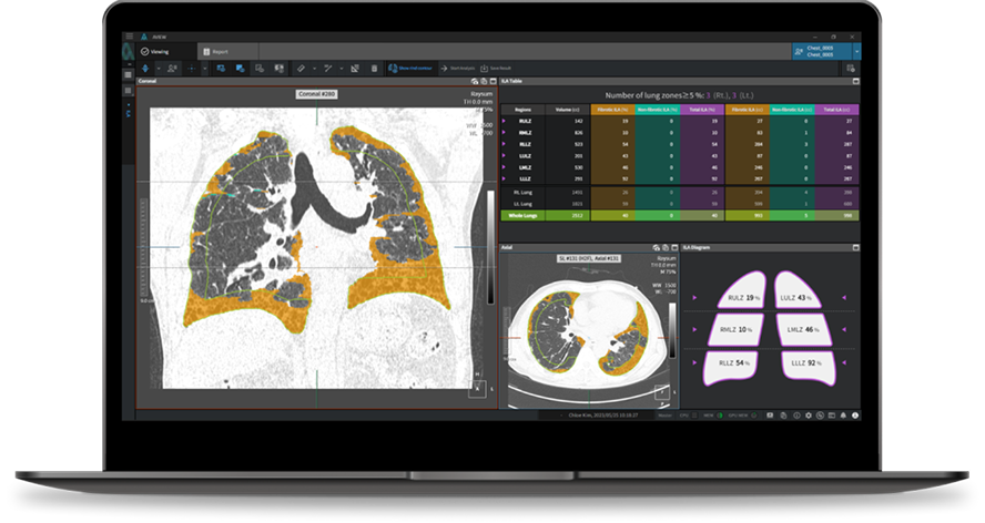
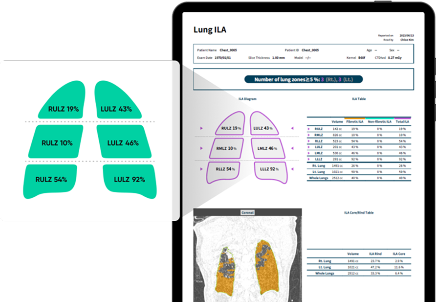
The analysis meticulously assesses lung segments to identify ILA, providing distinct evaluations for fibrotic and non-fibrotic types.
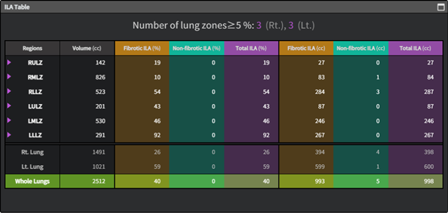
The system offers both quantitative and intuitive results. By dividing the lungs into six zones, it emphasizes areas where ILA distribution surpasses a 5% threshold.
Sensitivity
Specificity
AUROC
Chae KJ, Lim S, Seo JB, Hwang HJ, Choi H, Lynch D, Jin GY. Interstitial Lung Abnormalities at CT in the Korean National Lung Cancer Screening Program: Prevalence and Deep Learning-based Texture Analysis. Radiology. 2023 May;307(4):e222828.
AI Software designed to Analyze BoneTrauma on X-Rays
The First Multi-Agent System for Radiology Reporting
Your personal AI assistant in the battle against breast cancer.
Enhancing Diagnostics through Intelligent Image Analysis